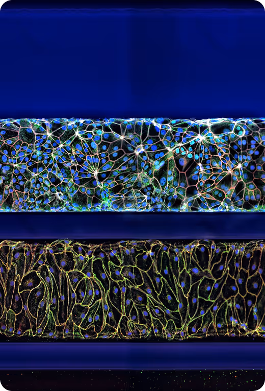
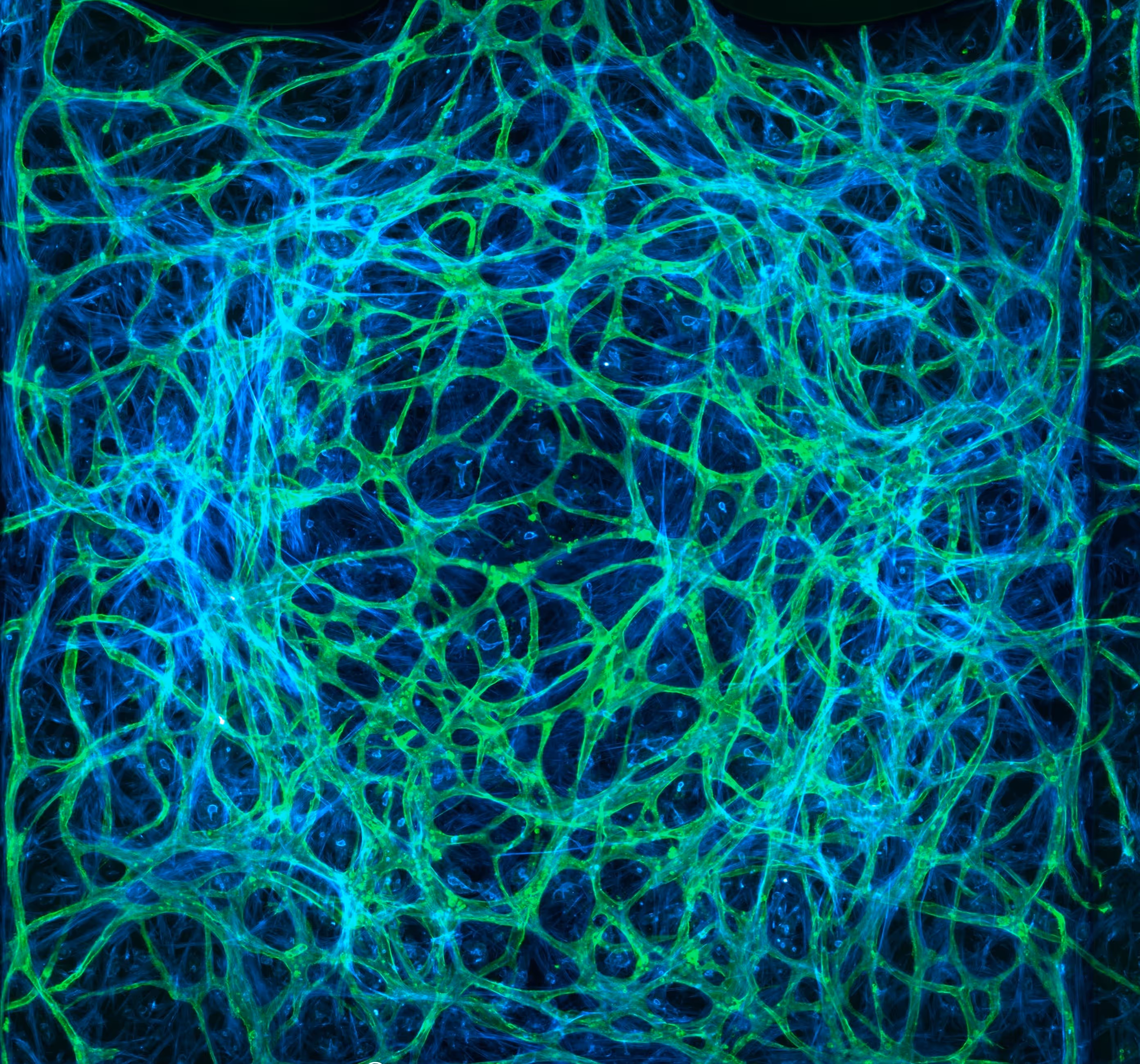
3D Vasculature Models
MIMETAS’ 3D vasculature models replicate (micro)vascular architecture under physiologically relevant flow conditions.
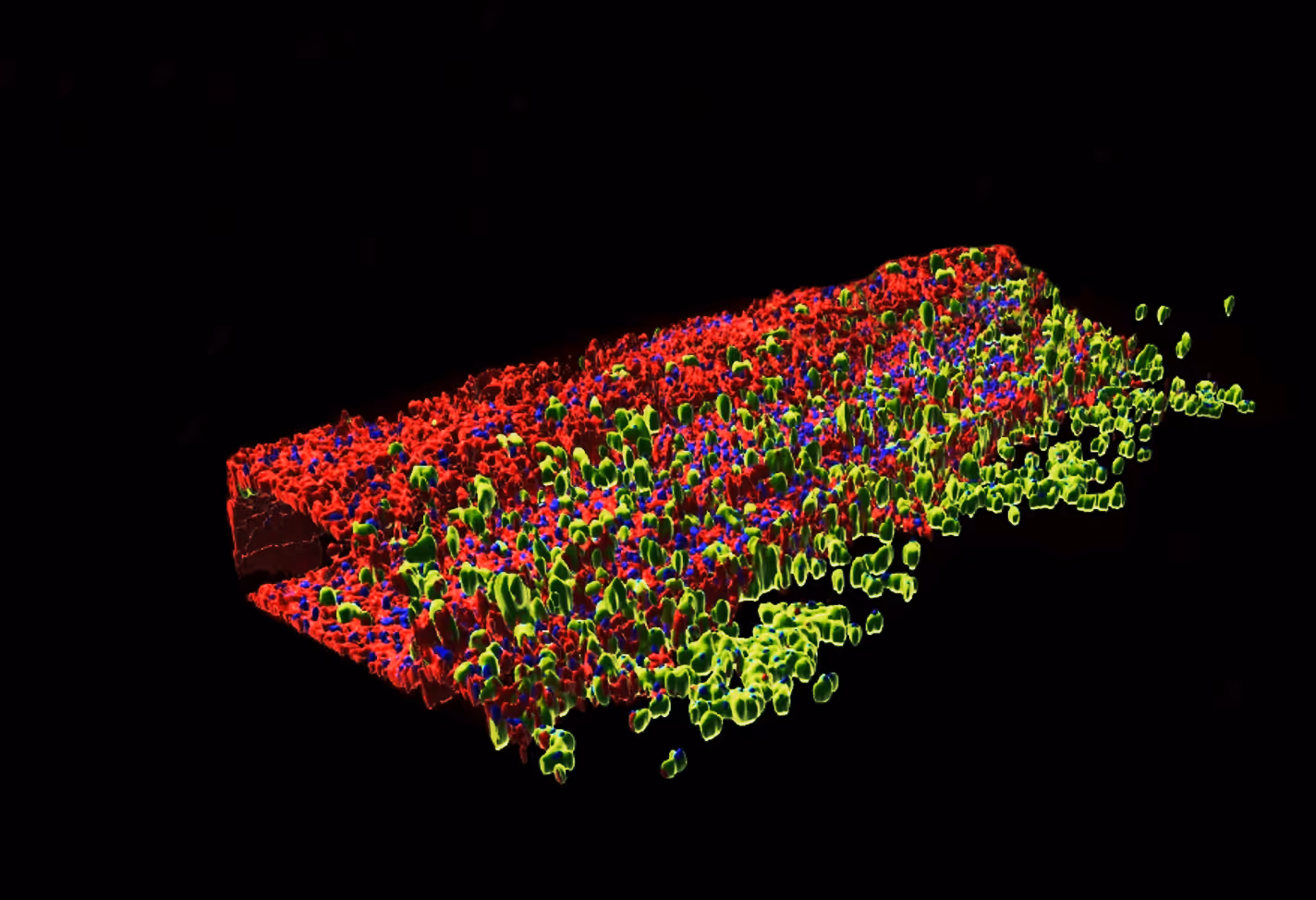
This supports insights into barrier function, immune cell trafficking and angiogenesis, which are key to evaluating drug efficacy and safety.
Vasculature Complexity Tailored to Your Research Question
MIMETAS vasculature models mimic the vascular microenvironment, including the endothelial barrier, extracellular matrix, and supporting cell types.
Available Model Configurations:
Organ-specific endothelial monocultures among which coronary artery, lymphatic, small intestine, glomerular and skin (micro)vasculature
Immune-inclusive co-cultures (PBMCs, monocytes, T cells)
Co-cultures (smooth muscle cells, pericytes, fibroblasts, tumor cells)
Vascular plexus (vascular bed)
A Range of Formats to Fit Your Needs
We are here to support you with bespoke services, ready-to-go solutions, or product support packages.
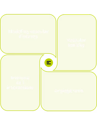
A Range of Applications
Disease modeling
Vascular toxicity
Inflammation
Hyper/hypo vascularization
Selected Resources

Publication
.avif)