


A scalable solution for 8 weeks microvasculature-on-a-chip with unidirectional flow
Microvasculature is not just a passive conduit. It actively regulates tissue homeostasis, barrier function, immune trafficking, and inflammatory signaling. As a result, vascular biology is central to both efficacy and safety questions across therapeutic areas.
Indrug discovery and development, vascular models are increasingly important for:
· Safety assessment: detecting compound-induced changes in barrier integrity, perfusability, and inflammatory activation
· Chronic exposure studies: capturing effects that emerge over weeks (e.g., progressive remodeling, delayed toxicity, cumulative stress)
· Mechanistic insight: linking endothelial–stromal interactions to functional outcomes
· NAMs readiness: supporting the shift toward New Approach Methodologies by providing more predictive, human-relevant data
However, many vascular microfluidic configurations described in the literature are difficult to reproduce at scale. Common barriers include limited standardization, nonstandard materials, complex hardware, and insufficient tissue maturation.
What’s unique about the MIMETAS approach
MIMETAS designed its microvasculature-on-a-chip solution around a practical requirement: make long-term, perfused vascular networks accessible in a standard plate workflow.
OrganoPlate® Graft UniFlow retains key OrganoPlate platform characteristics that lower the barrier to adoption:
· 384-well plate-based microfluidic format enabling high-throughput studies
· 32 independent chips per plate for parallelization and screening
· Membrane-free design to enable direct cell–ECM and cell–cell interactions
· Glass plate-based microfluidic titer plate built for imaging compatibility
· Use of low-absorbent materials to supportcompound testing
· Compatibility with microscopes, plate readers, and robotic handlers
The differentiating capability is the platform’s ability to maintain physiological unidirectional flow in a pump-free setup, supporting perfusion conditions that are more representative of in vivo vascular biology, while keeping operation simple.
How OrganoPlate® Graft UniFlow enables gravity-driven unidirectional flow
Each chip contains three functional elements:
· A perfusion channel
· A bypass channel
· A gel chamber with an opening used for seeding cells in an ECM hydrogel
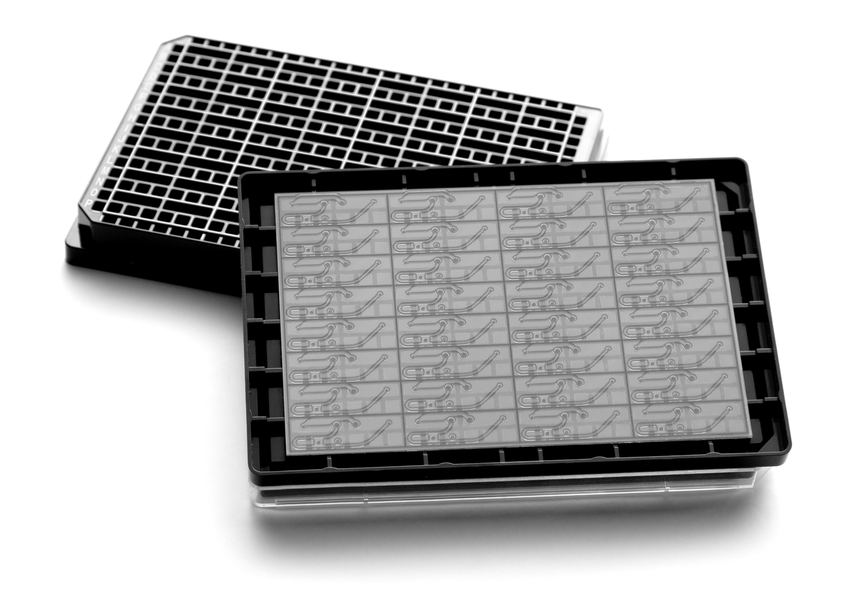
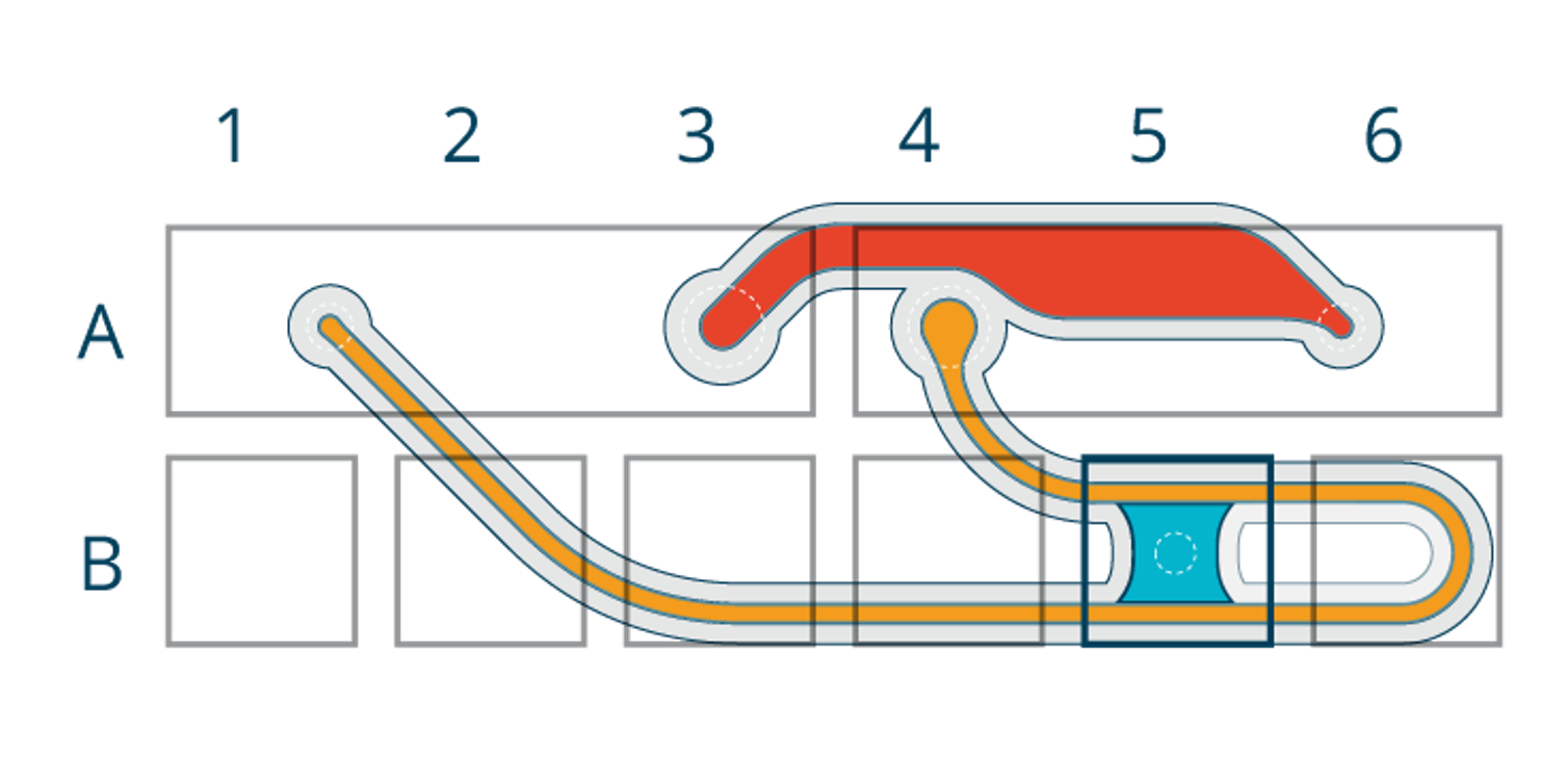
Unidirectional flow is generated through rocking that alternates between two phases:
1. Perfusion phase (~1 minute) When the plate rocks to one side, medium covers the opening that feeds the perfusion channel inlet. Medium enters the perfusion channel and flows through the microvascular network formed in the gel chamber toward the opposite long well. Any medium that does not pass through the network is routed through a loop in the perfusion channel, ending in the same destination well.
2. Reset phase (~20 seconds) When the plate rocks to the opposite side, medium covers only the opening corresponding to the bypass inlet. In this configuration, medium flows exclusively through the bypass, and no medium enters the perfusion channel, preventing backflow through the vascular network. After ~20 seconds, the plate returns to the perfusion orientation and the next cycle begins.
By separating perfusion from reset, the system achieves exclusively unidirectional flow through the microvascular network using gravity alone.
Evidence of robustness and long-term performance
MIMETAS´validation strategy was built around:
· Structural characterization (3D organization, endothelial markers)
· Robustness and reproducibility (low variability, minimal operator effect, transferability across labs)
· Fit-for-purpose pilot contexts of use (inflammation/immune responses and compound-induced vascular toxicity)
Akey capability for chronic studies is longevity: the blood vessel networks are reported to remain fully perfusable for at least 57 days, enabling experimental designs that extend beyond the typical 1–2 week window of many self-assembled microvascular models.
Case study: vascular remodeling and acute/chronic toxicity
A recent case study conducted by our researchers at MIMETAS demonstrates how a self-assembled, perfusable 3D microvascular network under unidirectional flow can be used for both acute and chronic vascular toxicity workflows. Our model is characterized by long-term perfusability (reported ≥57 days) alongside evidence of vascular maturation and remodeling over time, including preserved macromolecule retention (barrier integrity), changes in network morphology consistent with flow conditioning, and stromal reorganization toward perivascular stabilization. In pilot contexts of use, our platform supports quantification of inflammation-mediated immune responses (e.g., PBMC adhesion/extravasation dynamics that shift with vessel maturation) and compound-induced vascular toxicity, where readouts such as perfusability, permeability, stromal remodeling, and inflammatory markers reveal distinct response signatures across compounds and exposure windows.
How to access the platform: OrganoAccess and CRO services
To enable access to this microvasculature-on-a-chip solution we have recently launched OrganoAccess, an integrated offering designed to help researchers implement the technology in their own lab by combining access to the platform with enablement and support—so teams can move faster from setup to discovery.
OrganoAccess combines the OrganoPlate® Graft UniFlow platform with the practical enablement needed to implement it successfully, including validated cells and reagents, optimized protocols/SOPs for vascular network formation and downstream assays, and scientific support for troubleshooting and experimental design. The goal is to reduce the time spent on setup and optimization so teams can move faster into fit-for-purpose studies.
Forprojects requiring customized biology (e.g., immune co-culture, epithelial interfaces, patient-derived material, or tailored endpoints), MIMETAS CRO services can support development of fit-for-purpose models either in collaboration with your team or executed by MIMETAS experts.