Chat with us, powered by LiveChat
  • Get inspired by peer-reviewed publications of our scientists, partners, and customers around the globe.

  • Giving you some food for thought. Read our blogs to learn more about 3D tissue culture, research backgrounds, developments, and its future outlook.
  • Get inspired by research done by our scientists, partners, and customers around the globe.

  • Learn about our mission, vision, the history of the company, and find out what we mean with MIMETAS-do.
open menu icon close menu icon
EN

FAQ


Do you have any questions?
You might find here the answer you're looking for.

Filter on your topic of interest to get the related frequently asked questions.

All
All
Orders & Delivery
OrganoPlate Specifications
Gel loading
Cell culture
Perfusion
Assays
Imaging
Filter

OrganoPlate Specifications

Should I use an OrganoPlate® 2-lane or a 3-lane?

The main difference between the OrganoPlate® 2-lane or 3-lane is the extra channel in the 3-lane. This allows for more culture options such as: creating a gradient, having two types of ECM besides each other, having basal access to a tube, etc. This makes the OrganoPlate® 3-lane a necessary option for doing e.g. transport assays. The OrganoPlate® 2-lane however, is more fit for high-throughput screenings as it has 96 tissue culture chips per plate instead of the 40 tissue culture chips that the OrganoPlate® 3-lane provides.

Check out the OrganoPlate Family and find out which type fits your needs.

Do I need any special equipment for using OrganoPlate®?

The OrganoPlate® is an SBS-standard 384-well plate size, 127.76 x 85.48 x 14.8 mm (l x w x h) (17.3 mm height with lid) and is compatible with the standard laboratory systems (microscope, plate reader).

What material is the OrganoPlate made of?

  • Top plate: virgin polystyrene.
  • Bottom: optical quality 150 μm glass (#1 coverslip thickness)
  • Microfluidics: glass, proprietary polymers, biocompatible and low-compound-absorbing

What size is the OrganoPlate?

The OrganoPlate® is a SBS-standard 384-well plate size: 127.76 x 85.48 x 14.8 mm (l x w x h) (17.3 mm height with lid)

What are the sizes of the channels per type of OrganoPlate?

OrganoPlate® 2-lane:

  • Channel width:
    • Perfusion channel: 300 µm
    • PhaseGuideTM: 100 µm
    • ECM-gel channel (including PhaseGuideTM): 500 µm
  • Channels height: 220 µm

OrganoPlate® 3-lane 40:

  • Channel width:
    • Perfusion channels: 300 µm
    • PhaseGuideTM: 100 µm
    • ECM-gel channel (including both PhaseGuidesTM): 550 µm
  • Channels height: 220 µm

OrganoPlate® 3-lane 64:

  • Channel width:
    • Perfusion channels: 425 µm
    • PhaseGuideTM: 100 µm
    • ECM-gel channel (including both PhaseGuidesTM): 500 µm
  • Channels height: 220 µm

OrganoPlate® OrganoGraft:

  • Channel width:
    • Perfusion channels: 400 µm
    • PhaseGuideTM: 100 µm
    • ECM-gel channel (including both PhaseGuidesTM): 2000 µm
  • Channels height: 220 µm

You can find this and other product specifications in our Knowledge Center in the Brochures section.

Can I flip my OrganoPlate upside down?

Yes, because the medium in the OrganoPlate is in small wells, capillary forces ensure that the liquid does not leak out when you flip the OrganoPlate, even if there is medium in the OrganoPlate.

What are the recommended storage conditions for the OrganoPlate?

We recommend to store the OrganoPlate at 4°C upon arrival. The optimal performance, for ECM filling, can be achieved when the OrganoPlate is used within 12 months.

Can I freeze my samples within the OrganoPlate®?

Empty OrganoPlate® or OrganoPlate® cultures with or without cells, should never be stored in the freezer or frozen down. Freezing of empty plates will cause delamination of the glass bottom from the microfluidics. If water is present in the channels, its expansion will also mechanically disrupt the microfluidics leading to irreversible loss of the sample due to contamination across the different chips. If you want to store your plate post-fixation or post-staining, store the plate at room temperature for short-term or at 4°C for longer storage.

Is the OrganoPlate® flatness compatible with my microscope auto-focus feature?

The OrganoPlate® is optimized for high content screening application using standard auto-focus methods. The OrganoPlate® has an inter- and intra-chip bottom flatness within 120 μm and 5 μm, respectively. Excellent optical performance is ensured by the 1H coverslip glass equivalent plate bottom thickness of 150 +/- 5 μm. For optimal performance we advise performing laser based autofocus for each chip by focussing on the outside bottom surface of the glass and offsetting by the glass thickness.

Orders & Delivery

How do I order products through the online quote form?

You can fill out the quote form on our website and the next working day our Sales Team will reach out to you with an official quote that meets your request. Once you accept our quote, our Sales Team will guide you further on how you can proceed with ordering.

How will my order be delivered?

We make efforts to deliver our products within 4 (four) weeks after the agreement takes effect. Shipping and delivery is handled by FedEx or YSDS. Find our Terms & Conditions here.

Can my order be delivered to a different address then my billing address?

Yes, you can always arrange for your order to be delivered to a different address within the same country. You can specify the billing address and delivery address with our Sales Team.

Can my order be delivered to a PO Box address?

Unfortunately, this is not possible.

To which countries does MIMETAS ship orders?

We deliver the OrganoPlate®, OrganoReady® Collagen, OrganoFlow, and OrganoTEER® products worldwide.
The cell based OrganoReady products are available within Europe, United Kingdom, Japan, United States and Canada

How can I cancel an order?

Once an order has been placed, it is final and, unfortunately, cannot be canceled. If you no longer want the items, you can send them back as soon as you receive them (within 14 days). You will then be refunded once we have received the items in the original state of the package, unopened and undamaged.

I haven’t received my order, what should I do?

If you have not received the items you ordered within 4 (four) weeks, please contact our Customer Service department through support[at]mimetas.com

I received an item I did not order, what should I do?

If you have received an item you did not order, please contact us through support[at]mimetas.com and describe the products that you did receive.

Gel loading

Which ECMs / gels work in the OrganoPlate®?

The OrganoPlate® is compatible with a range of ECM gels that can transition from a liquid to a solid-state. These gels include, but are not limited to collagen-I, Matrigel®, Matrigel®-growth factor reduced BME, and HyStem gels.

How much gel do I need?

Depending on the OrganoPlate you work with and gel of choice, you need 1-2 µL of gel per tissue culture chip. Make sure to prepare a 40% excess to compensate for loss of gel due to the dead volume of the repeating pipet. For the preparation of collagen-I gel, prepare at least 100 µL total volume to ensure proper mixing of all components of the gel.

Is it possible to coat the OrganoPlate?

The materials that make up the OrganoPlate tissue culture chips are compatible with cell culture. However, certain cell types benefit from or require the use of a coating with extracellular matrix proteins, as they also do in other culture vessels. Coating of the channels of the OrganoPlate's tissue culture chips helps to improve cell attachment, tubule formation, or biological performance of certain cell types.

How long should I incubate my gel?

The incubation time depends on your gel of choice. We advise incubating collagen-I and Matrigel® for 15-20 minutes at 37°C before adding culture medium. After incubation, the addition of medium in the adjacent medium channel will ensure the gel will not dry out. If you plan to seed tubes, add 20 µL of PBS or HBSS to the gel inlet well after gelation occurred and place in an incubator overnight before seeding the tubular cells. The PBS or HBSS should be removed from the gel inlet before you seed your cells.

What does a properly seeded gel channel look like?

After seeding your gel in the gel channel, that channel is observed as dark when you look at the bottom of the plate (you can flip the plate upside down without worrying about liquids flowing out). The adjacent medium channel(s) are still empty and observed as transparent.

When looking from the top, a thin, dark, horizontal line can be observed in the observation window, indicating your gel has filled properly. This dark line is also observed when you look at the chip under the microscope.

Why does my gel not fill the channel all the way to the end?

Improper filling of the gel channel can occur if you are working with a very viscous gel or if your gel is already partly polymerized. When working with Matrigel® or Matrigel®-like gels, thaw the gel according to the manufacturer’s instructions and make sure the gel is kept on ice continuously to prevent it from polymerizing. Using pre-cooled Eppendorf tips will also prevent this.

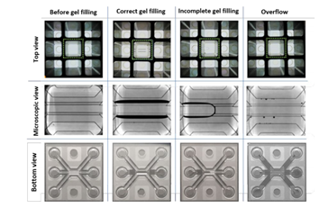
If your gel does not fill the gel channel properly, try increasing the seeding volume. For example, for OrganoPlate® 2-lane and 3-lane with a 400µm channel width, the range of collagen-I gel volume to seed varies between 1.5-2.1 µL. We advise starting with a seeding volume of 2 µL. If the gel does not properly fill the channel all the way, increase the seeding volume until you find the volume that works for your experiment. The Sartorius eLINE electronic repeating pipette (#735021) is very well suited for this purpose.

Why does my gel also fill the medium channel(s)?

If your gel fills both channels, you are unable to seed cells in the medium channel or to use that channel for perfusion. If you seed correctly - see our instruction manuals on how to correctly seed - overflow from the gel channel into an adjacent medium channel can be solved by lowering the gel seeding volume. The perfect seeding volume is influenced by the viscosity of the gel and the temperature in your lab. For example, for OrganoPlates® 2-lane and 3-lane with a 400µm channel width, the range of collagen-I gel volume to seed varies between 1.5-2.1 µL. We advise starting with a seeding volume of 2 µL. If overflow occurs, lower the seeding volume until you find the volume that works for your experiment. The Sartorius eLINE electronic repeating pipette (#735021) is very well suited for this purpose.

Furthermore, retain from touching the glass bottom directly. This could also lead to overflow.

What is the dark line / shadow that I observe after gel seeding?

When the gel flows into the gel channel, the PhaseGuide™ (a small rim that functions as a pressure barrier) prevents it from flowing into the adjacent medium channel(s). After filling the channel, the gel stretches partially into the next channel, which can be observed as a shadow or a dark line in the medium channel. When you see the dark line, it is a sign you’ve seeded the gel properly. Once you have added medium to the adjacent channel this dark line should have disappeared. If it remains it means there is air in the medium channel. You can read all about the physics behind the PhaseGuide™ and meniscus stretching in our publication.

What pipette should I use?

Any pipette that can dispense volumes in the 0.5-2.5 µL range can be used. However, if you are experiencing difficulties with gel loading and cell seeding, we recommend using the Sartorius eLINE® electronic pipette (Sartorius, #735021 for accurate dispensation of volumes ranging from 0.2 to 10 µL or the Eppendorf® Multipette® M4 with the Eppendorf® Biopur® 0.1 mL tip (VWR #613- 2067) for dispensation of 2 µL.

Cell culture

How can cell invasion in ECM gel be avoided?

In case of undesired cell invasion into the gel, the use of MMP inhibitors is recommended (e.g. addition of 10 μM of MMP-I inhibitor GM6001 (Abcam, ab120845) to the culture medium).

How many cells do I need?

You need 2 µL of cell suspension per tissue culture chip. The cell density of the suspension needs to be optimized for your specific culture but generally ranges from 5-30*103 cells/µL.

What cells have been tested in the OrganoPlate?

OrganoPlate® users have worked with a wide range of cells, both human and animal-derived. Primary cells, (cancer) cell lines, induced pluripotent stem cells, and organoid (derived) cells have successfully been cultured in the OrganoPlate® for among others vascular, kidney, gut, liver, CNS, and cancer models. Check our Knowledge Center for all our publications.

How do I change the medium in the OrganoPlate?

A medium change can be done by aspirating all the media from the medium inlet and outlet wells and replacing it with fresh culture medium. A mechanical single channel or multichannel aspirator can be used. Aspiration will not disturb the cells in the microfluidic channels. Place the tip(s) of the aspirator in the corner of the well while aspirating as you would do with a regular culture plate and remove all liquid. Fresh culture medium can then be added by manual or automatic pipetting into the medium inlet and outlet wells. Watch this Medium change video or read our protocol.

How often do I have to change the medium?

For most cells, we advise changing the medium 2-3 times a week (for example on Monday, Wednesday, and Friday). If your cells are highly metabolically active, you can consider changing the media more often.

Can I reuse my OrganoPlate after washing?

We do not recommend reusing chips in a plate, as we cannot guarantee the quality when reusing it.

How long can the cells be cultured in OrganoPlate?

It will depend on your cell type and your seeding density. For example, we have been able to keep different gut cells, such as Caco-2, in culture for up to a month.

How do I mitigate against edge-effect within an OrganoPlate?

We recommend randomizing the experimental conditions across all the chips in the OrganoPlate. If media evaporation in the external chips is affecting your results, especially with long term cultures, we recommend substituting the regular microplate lids, with hydrated MicroClime® Microplate Beckman Coulter 0015717. When imaging, substitute the MicroClime® lids with the original or a sterile plate lid like High Profile Greiner Bio Clear PS Lids (art. 656101) or equivalent. Lids can remain on the plate when imaging using fluorescence inverted microscopy.

Perfusion

Do I need an OrganoFlow®?

Except for neuronal cultures, all our tissue culture models (i.e. liver, gut, kidney, vasculature, cancer, etc.) were shown to benefit strongly from medium perfusion. Perfused cultures show improved cell viability, the polarization of tubular structures, and upregulation of relevant transporters.

If you first want to try the OrganoPlate before purchasing a rocker, you can use a regular interval rocker for your first experiments. We’ve observed the formation of tubular structures of HUVECs using a regular interval rocker. However, to obtain optimal barrier formation and allow more controlled flow profiles, the OrganoFlow® is advised.

What flow speeds am I working with?

An overview of flow speed and shear stress is available upon request. Please contact support[at]mimetas.com.

Which cell types can grow without perfusion?

Neuronal cultures show optimal viability when cultured without perfusion. This includes both long-term differentiation of neural stem cells and short-term cultures of already differentiated neurons.

Is the flow bidirectional or unidirectional?

The flow is bidirectional. The OrganoPlate is placed on an OrganoFlow. A flow is created by passive liquid leveling as the medium flows from medium inlet to outlet and vice versa. This allows you to create differentiated/polarized tissues without the hassle of pumps or tubes.

What rocker settings should I use?

We observed that a 7° inclination and an 8-minute interval work well for most culture types grown in the OrganoPlate® 2 lane or 3 lane 40. Cultures grown in the newer OrganoPlate® 3-lane 64 or Graft Plate will benefit from an equivalent inclination of 14° and 8-minute interval.

Is it possible to control the flow?

Yes, you can control the flow by changing the inclination angle of the rocker as well as by changing the rocker’s interval.

Imaging

Is the OrganoPlate suitable for imaging?

Yes, the OrganoPlate is excellent for imaging purposes. The bottom of the plate is made of high optical quality 150 μm glass coverslip-thickness glass, allowing unparalleled optical read-outs. The OrganoPlate’s microtiter plate footprint makes it compatible with standard laboratory imaging systems.

What types of imaging can I do on the OrganoPlate?

The OrganoPlate is compatible with regular (time-lapse) phase contrast and fluorescent imaging as well as confocal imaging. The OrganoPlate is not compatible with electron microscopy.

Do I need a special microscope?

No, the OrganoPlate’s microtiter plate footprint makes it compatible with standard laboratory imaging systems.

Assays

Can I perform cell viability assays?

Yes, the OrganoPlate is compatible with various cell viability assays that employ fluorescent, luminescent, or absorbance read-outs. These assays include but are not limited to, the LIVE/DEAD® viability assay, the CellTiter-GLO™ assay, and the Alamar Blue assay.

Can I perform an immunostaining?

Yes, the OrganoPlate is ideal for immunostaining and imaging. Our immunostaining protocol can be found here. The plate is compatible with common fixation methods, such as formaldehyde and methanol fixation, and we do not observe problems with antibodies permeating the gel. If you prefer using your lab’s specific immunostaining protocol, this will very likely translate to OrganoPlate cultures as well. However, longer incubation times can be necessary compared to standard protocol (comparable to staining tissue slices).

Can I measure the integrity of my barrier tissue?

Yes, we have developed robust protocols to assess the integrity of barrier tissues such as endothelial and epithelial tubes. The protocol of our barrier integrity assay for OrganoPlate® 2-lane and 3-lane can be found here.

Can I isolate RNA and DNA from OrganoPlate cultures?

Yes, you can successfully isolate high-quality RNA or DNA from the OrganoPlate using traditional Trizol® extraction or column-based isolation kits. Depending on the nature of your culture and the amount of RNA or DNA needed, pooling the contents of several chips may be necessary to obtain sufficient material. To give you an estimate: from an endothelial tube in one chip of an OrganoPlate® 3-lane , we typically collect 50 nanograms of RNA.

Can I sample from the OrganoPlate?

Yes, samples can be easily collected by taking the medium from the chip. This medium can be used to analyze the contents by various assays and read-outs, such as ELISA and mass spectrometry. In the OrganoPlate® 3-lane, you have access to both the apical and basal sides of the chip.

Can I isolate live cells from the OrganoPlate?

Yes, if you want to remove cells from the chips (for i.e. flow cytometry applications) you can. For instance, by trypsinizing your cells.

Cookies

‘May we use cookies?
Hi there! Thanks for visiting our website. We use cookies to keep track of our website statistics to optimize the user experience. We also use cookies for marketing purposes. You can set your preferences by selecting the options below. Terms of Use & Privacy Policy

Accept all
Accept selected
Decline all