• Get inspired by peer-reviewed publications of our scientists, partners, and customers around the globe.

  • Giving you some food for thought. Read our blogs to learn more about 3D tissue culture, research backgrounds, developments, and its future outlook.
  • Get inspired by research done by our scientists, partners, and customers around the globe.

  • Learn about our mission, vision, the history of the company, and find out what we mean with MIMETAS-do.
open menu icon close menu icon
EN

Perfused 3D Angiogenic Sprouting In A High-Throughput In Vitro Platform

On-Demand Webinar

Studying angiogenesis in a manner that is representative of the in vivo situation can be a challenging task. Crucial elements in modeling angiogenesis are the formation of 3-dimensional sprouts having a lumen and clearly defined tip and stalk cells. Other important aspects include the presence of an extracellular matrix and the perfusion of blood vessels. What if there was a way to study angiogenesis in vitro in a way that would allow you to model all these aspects of angiogenesis in an easy and quick way.

  • You could not only study the onset of angiogenesis, but also key aspects that occur during the maturation phase.
  • You would be able to access the lumina of your angiogenic sprouts, and perfuse them too.
  • Would you like to apply a stable gradient of growth factors to direct the formation of capillaries?


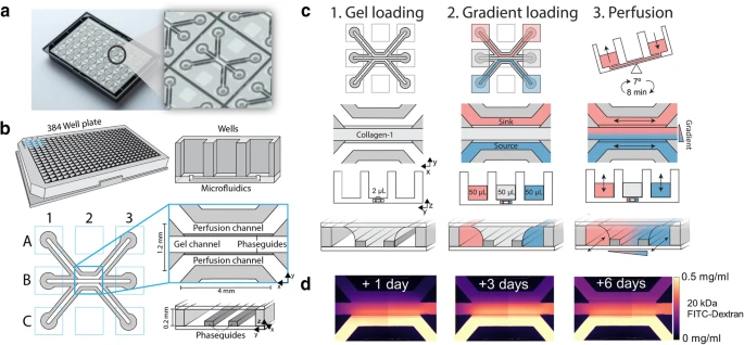
Think about all the possibilities that would open up, if you had such a system. In this webinar, you will learn about a robust microfluidic platform (OrganoPlate®) that will allow you to do all of the above. The OrganoPlate® platform is already successfully being used by the top 10 pharma as well as by hundreds of academic researchers worldwide. Unlike most microfluidic devices used today, the OrganoPlate® does not make use of any tubings nor pumps. As a matter of fact, it looks and handles exactly like a standard 384-wells plate. This means that it is compatible with all your equipment. In this webinar, you will learn how to work with collagen gel, form a blood vessel, applying a perfusion flow and creating a gradient of angiogenic factors so that you can study the onset of angiogenesis, as well as key aspects that occur during the maturation phase.


In this webinar, you will learn:

  • How to study the initiation and progression of angiogenic sprout formation
  • How to build perfusable 3D microvessels
  • How to create a stable and reproducible gradient for your sprouting angiogenesis studies
  • How to easily assess the permeability and integrity of the lumen and gauge your model leak-tightness

Register Here


What others have said about the OrganoPlate®

“With the MIMETAS OrganoPlate, we developed a 3D brain-on-a-chip model that made easy to do high-throughput and high-content imaging to evaluate the acute toxicity of neurotoxins. OrganoPlate allowed the study of membrane-free vascular formation in a dynamic environment and further provided reliable and repeatable experiments with strong imagining capability.” — Dr. Yeoheung Yun, Associate Professor at North Carolina A&T State University

“Often you have to compromise: it’s either the throughput or it’s the complexity of the model. Getting both in the same platform…no other platforms can do that!” – Senior Scientist from top pharma


Speaker

Vincent van Duinen, Scientific Project Lead at MIMETAS
Vincent obtained his PhD in 2013 at the group of Analytical BioSciences at Leiden University, under the supervision of Thomas Hankemeier and Paul Vulto. In his thesis titled 'Microvasculature in Microfluidics', he showed his pioneering work on the development of vascular models systems using the MIMETAS OrganoPlate. His post-doctoral research was performed at the Leiden University Medical Center, where he focused on the usage of clinical samples to bridge the gap between in vitro and in vivo using microvasculature-on-chip. At MIMETAS, Vincent leads projects on Liver fibrosis, with a strong emphasis on functional liver vasculature. This will aid in the discovery of novel disease pathways that drive liver fibrosis and steatosis.


Related publications


Cookies

‘May we use cookies?
Hi there! Thanks for visiting our website. We use cookies to keep track of our website statistics to optimize the user experience. We also use cookies for marketing purposes. You can set your preferences by selecting the options below. Terms of Use & Privacy Policy

Accept all
Accept selected
Decline all