On-demand Webinar
Key tissue modalities, such as vasculature and cell migration,  play an important role in maintaining homeostasis in multicellular tissues. For example, immune responses require the coordinated movement of cells by relying on chemotaxis, cell signaling, and cell-matrix interactions.
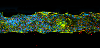
To analyze biological mechanisms of varying cell modalities in complex in vitro models, imaging techniques are pivotal for detailed cellular visualization. By using high-content screeners and laser-point scanning microscopes, researchers can visualize, characterize, analyze, and describe various tissue and disease phenotypes. Key assays of applications such as immune migration include barrier transmigration, T-cell patrolling, protein marker expression and associated changes in tissue for both healthy and diseases states.
Watch this webinar and join our speakers Thomas Olivier (MIMETAS) and Rouven Schoppmeyer, PhD (Nikon BioImaging Lab), as they present an overview of the benefits and hurdles of the latest imaging techniques utilized in advanced 3D tissue and disease modeling.
In this webinar, you will:
- Learn about advanced imaging techniques and applications used to study complex in vitro tissue and disease models
- Discover the OrganoPlate® platform’s compatibility with imaging techniques and related assays, and how it can further facilitate your research to obtain humanized data
- Explore key assays used for cell modalities such as immune migration
Access the recording here
Speakers
Thomas Olivier  is Lead Bioinformatician at MIMETAS. He received his BSc in Bioinformatics from the Hogeschool Leiden and started working at MIMETAS shortly afterwards as a Bioinformatics Engineer. In his current role, he and his team focus on image analysis, RNA-sequencing analysis and provide data-science services to the R&D team.
 Rouven Schoppmeyer, PhD is a Scientist at Nikon BioImaging Lab (NBIL). During his bachelor's and master's he studied Applied Biology and Biomedical Sciences in Rheinbach, Germany. In 2017, he received his PhD from Saarland University, during which he focused on 3D migration of lymphocytes and cell-mediated cytotoxicity using advanced microscopy techniques. After working at Sanquin/AMC Amsterdam to study transendothelial migration of human leukocytes, at NBIL he provides CRO services to academia and the private sector, specialized in complex functional assays, with a current focus on immune cell biology and neuroscience.
