Chat with us, powered by LiveChat
  • Technology
  • Services
    • Drug Development Partnerships
      • Expand your drug discovery capacity, shoulder to shoulder with our scientific team. Proprietary human disease biology in the OrganoPlate® platform. Together we make the therapeutics of tomorrow.
    • Custom Model & Assay Development
      • We create novel human tissue and disease models in the OrganoPlate® platform. Our experts are looking forward to developing assays according to your specifications.
    • Compound profiling & Screening
      • Get robust compound data in human tissue models through our services. With proven pheno­typic assays in the OrganoPlate® platform, we support your drug discovery and development needs.
    • All Services
      • We offer several services to support your drug discovery and development needs. Find the overview here.
  • Products
  • Applications
    • Perfused Tubules
      • Layered tissues with perfused tubules in the absence of artificial membranes form the heart of our permeability and transport science. Study cell interactions, permeability, absorption, transport, and transcytosis without physical barriers.

    • Cell-Cell Interactions
      • Co-culture layered & structured tissues without artificial membranes with perfect imaging, to study barrier-free cellular interactions, cell-cell signaling, and migration.

    • 3D Cell Migration
      • Evaluate the effect of chemotactic triggers or cells on the migration of cells through an extracellular matrix.

    • Angiogenesis
      • Membrane-free microvascular formation and growth through an extracellular matrix (ECM).

    • Vascularization
      • The missing link in tissue culture: add perfusable human vasculature to your tissue models, and recreate sophisticated microenvironments with OrganoPlate® Graft.

    • All Applications
      • OrganoPlate® enables you to study relevant 3D tissue biology by incorporating perfused tubules, co-culture, and full control over the tissue microenvironment. Find the overview of applications here.

  • Resources & Support
    • Knowledge Center
      • Visit our Knowledge Center to get up to speed with 3D tissue culture and to learn how OrganoPlate® supports your research needs.

        Read our publications, application notes, watch our webinars, or check out the supporting protocols and brochures. All compiled for you, by our scientists.

    • Publications
      • Get inspired by peer-reviewed publications of our scientists, partners, and customers around the globe.

    • Case Studies
      • Get inspired by research done by our scientists, partners, and customers around the globe.

    • Blogs
      • Giving you some food for thought. Read our blogs to learn more about 3D tissue culture, research backgrounds, developments, and its future outlook.
    • Workshops & Trainings
      • Kickstart your experiments with the most sophisticated 3D tissue culture platform. Find out which training fits your 3D tissue modeling needs best.
    • Technical Support
      • Any support questions about purchasing, products, or 3D tissue culture and analysis? Get in touch with our experts.
    • Request a quote
      • Thinking of using OrganoPlate for your research? Request a quote for the product(s) of your interest.

    • FAQ
      • Frequently asked questions answered by our experts. Find here the answer you're looking for.
  • Knowledge Center
  • About Mimetas
  • Expand your drug discovery capacity, shoulder to shoulder with our scientific team. Proprietary human disease biology in the OrganoPlate® platform. Together we make the therapeutics of tomorrow.
  • We create novel human tissue and disease models in the OrganoPlate® platform. Our experts are looking forward to developing assays according to your specifications.
  • Get robust compound data in human tissue models through our services. With proven pheno­typic assays in the OrganoPlate® platform, we support your drug discovery and development needs.
  • We offer several services to support your drug discovery and development needs. Find the overview here.
  • Start today with 3D tissue modeling
    Kickstart your first experiments with the OrganoPlate® through our OrganoStart packages.
  • OrganoFlow®
    Driving precisely-controlled perfusion flow in the OrganoPlate® platform

  • OrganoTEER®
    Enabling fast, automated, and impedance-based TEER measurements in OrganoPlate®. Assay 40 or 64 tissue culture chips with a few clicks, in less than two minutes.

  • Start today with 3D tissue modeling
    Kickstart your experiments with the most sophisticated 3D tissue culture platform. Find out which training fits your 3D tissue modeling needs best.
  • OrganoPlate® is the solution for all in vitro tissue culture applications. Explore the entire OrganoPlate® family and its dedicated instruments here.

  • Thinking of using OrganoPlate for your research? Request a quote for the product(s) of your interest.

  • Layered tissues with perfused tubules in the absence of artificial membranes form the heart of our permeability and transport science. Study cell interactions, permeability, absorption, transport, and transcytosis without physical barriers.

  • Co-culture layered & structured tissues without artificial membranes with perfect imaging, to study barrier-free cellular interactions, cell-cell signaling, and migration.

  • Evaluate the effect of chemotactic triggers or cells on the migration of cells through an extracellular matrix.

  • Membrane-free microvascular formation and growth through an extracellular matrix (ECM).

  • The missing link in tissue culture: add perfusable human vasculature to your tissue models, and recreate sophisticated microenvironments with OrganoPlate® Graft.

  • OrganoPlate® enables you to study relevant 3D tissue biology by incorporating perfused tubules, co-culture, and full control over the tissue microenvironment. Find the overview of applications here.

  • Visit our Knowledge Center to get up to speed with 3D tissue culture and to learn how OrganoPlate® supports your research needs.

    Read our publications, application notes, watch our webinars, or check out the supporting protocols and brochures. All compiled for you, by our scientists.

  • Get inspired by peer-reviewed publications of our scientists, partners, and customers around the globe.

  • Get inspired by research done by our scientists, partners, and customers around the globe.

  • Giving you some food for thought. Read our blogs to learn more about 3D tissue culture, research backgrounds, developments, and its future outlook.
  • Kickstart your experiments with the most sophisticated 3D tissue culture platform. Find out which training fits your 3D tissue modeling needs best.
  • Any support questions about purchasing, products, or 3D tissue culture and analysis? Get in touch with our experts.
  • Thinking of using OrganoPlate for your research? Request a quote for the product(s) of your interest.

  • Frequently asked questions answered by our experts. Find here the answer you're looking for.
  • Learn about our mission, vision, the history of the company, and find out what we mean with MIMETAS-do.
EN

Modelling and prevention of acute kidney injury

Leiden, February 24, 2022 – Scientists from MIMETAS and Astellas introduce a combined human renal proximal tubule/blood vessel-on-a-chip model in the OrganoPlate® 3-lane 40 to study acute kidney injury (AKI) caused by renal ischemia/reperfusion injury (rIRI). This article has just been published in Kidney360.

The kidneys are responsible for removing waste products from our blood, while simultaneously facilitating the reuptake of essential nutrients. This results in a high energy demand, and dependency on supply of oxygen and nutrients through the blood flow. Both disruption (ischemia) and the restart (reperfusion) of the blood flow can cause a loss of function of kidney cells and ultimately lead to AKI. This results in over 13 million yearly diagnoses, where a person’s kidneys suddenly shut down, causing toxins to accumulate in the bloodstream. AKI progression can lead to chronic kidney injury, cardiovascular pathology, and ultimately death.

To study the systemic symptoms of AKI, in vivo animal models are often used. Unfortunately, due to interspecies differences, not all drug candidates that shown efficacy in the animal models translate to efficacious candidates in clinical trials. Similarly, traditional 2D in vitro models fail to recapitulate the kidney structure and function, which makes them unsuited to predict clinical outcomes. Organ-on-a-chip technology offers a physiologically relevant alternative by culturing in vitro models in 3D to mimic the structure and function of the kidney, increasing the predictive value required for drug development and disease research.

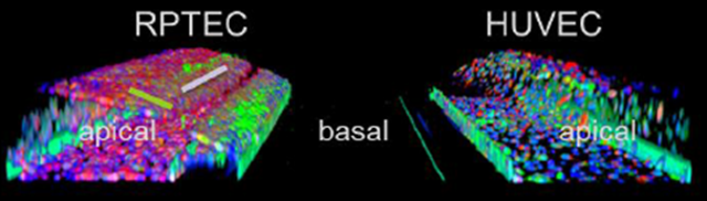
In this publication, an advanced kidney model was developed to study rIRI induced AKI. This model is based on a perfused 3D proximal kidney tubule in addition to a 3D perfused blood vessel, cultured from RPTEC and HUVEC cells, respectively. In this model, rIRI could be captured by altering oxygen levels, nutrient availability, and perfusion flow. It was shown that a combination of low oxygen, reduced glucose, and interrupted flow was potent to significantly damage the kidney tubules. Additionally, the damaging effect was amplified upon reperfusion of the tubules and could be alleviated through an adenosine treatment. The robustness and high-throughput capabilities of the model make it a valuable asset to advance pathophysiological research and drug development against AKI.

Go to the publication Sign up for our Newsletter

Cookies

‘May we use cookies?
Hi there! Thanks for visiting our website. We use cookies to keep track of our website statistics to optimize the user experience. We also use cookies for marketing purposes. You can set your preferences by selecting the options below. Terms of Use & Privacy Policy

Accept all
Accept selected
Decline all