Intestinal permeability research models are critical to deepening our understanding of barrier function of the intestinal epithelium, and associated pathologies.
To validate targets and test compounds efficiently, intestinal permeability platforms should meet several criteria. The ideal platform would:
- Be easy to use
- Remove the need for dedicated equipment
- Mimic inflammatory characteristics found in vivo
- Be robust, reproducible, and relatively fast
- Allow compound screening
Here, we describe the limitations of current traditional membrane inserts such as Transwells, and highlight the advantages of MIMETAS' OrganoPlate®, a platform that meets the above criteria and allows intestinal permeability models to be created in
fewer than five days.
Limitations of conventional permeable filter support systems
Conditions are static
Permeability models based on traditional membrane inserts typically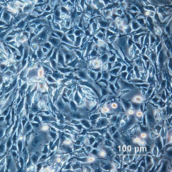 cultivate cells on a rigid membrane that separates two medium-containing compartments. A gradient is generated across the two chambers which allows cells to migrate through the porous membrane. However, mechanical forces are absent. Mild mechanical forces from shear stress are known to elicit biologically relevant signals in vivo, and intestinal epithelial cells are no exception. For example, one study found that apical shear stress is transduced by mechanosensitive microvillar protrusions to impact on an autophagic trafficking pathway.1 Models that neglect to provide shear stress are missing out on an important physiological component.
Extracellular matrix is absent
The extracellular matrix is another critical and complex aspect of in vivo physiology, and its absence has been shown to influence cell function, signaling, and adhesion, which directs cellular phenotype. A shift in the field towards 3D cell culture models have led to the development of extracellular matrix gels (formed from both synthetic and natural materials) to meet this demand.2
Cells are grown in a 2D monolayer
It is now accepted that 2D cultures do not necessarily reflect complex microenvironments found in vivo. Their limitations are particularly pertinent when it comes to disease-specific modeling, and this discrepancy is thought to be a significant contributor to the high failure rate in drug discovery.3 Cells in 2D culture experiments are heavily influenced by the stiffness of hard glass or plastic surfaces and show altered adhesion and migration characteristics.3 The stiffness of 3D extracellular matrices is so influential on cell migration, that bioengineers are hoping to create optimized ECM materials that help recruit repair cells to wound sites.4
Meanwhile, 2D platforms do not offer such tunable options, while altering cellular phenotype, gene expression, and biochemistry.5,6 More relevant models are needed to better predict the success of therapeutics.
Time to differentiation and assay readout time
The culturing and differentiation of Caco-2 (human colon adenocarcinoma) cells in Transwell inserts takes weeks, and most studies of Caco-2 cells allow 21 days for cells to form a differentiated monolayer before commencing experiments.7-9 Paracellular permeability assays, which are used to predict compound absorption across tight junctions between human intestinal epithelial cells, take approximately five hours.10
Accelerate and improve your permeability experiments with OrganoPlate
The OrganoPlate is an organ-on-a-chip platform that supports the culture of Caco-2 cells in a
tubular formation. Independent compartments can contain perfused leak-tight tubular tissue, enabling access to both the apical and basal sides. The OrganoPlate offers major advantages over conventional Transwell systems:
Intestinal permeability studies can be started four days post-seeding
In the time it takes to wait for cells to differentiate in a Transwell insert, you probably could have drafted a manuscript. In contrast to the 21-day culture period required in Transwell systems, Trietsch et al. (2017) report in Nature Communications the successful growth of leak-tight Caco-2 cells in tube formation in just 4 days.11 This is achieved via the intricate multi-channel design of the OrganoPlate. Once an ECM gel is introduced to the OrganoPlate platform, epithelial cells can be seeded and sediment directly against the ECM.
Upon the application of flow, cells proliferate and line all surfaces of the perfusion channel, forming a confluent tubular structure. The recommended time at which you can begin your permeability tests coincides with optimal barrier function, four days post-seeding.11 Additional time is also saved, due to the increased sample numbers within a plate; the OrganoPlate 3-lane 40 supports up to 40 tissue models on a single plate. Paracellular permeability readouts can be obtained in 15 minutes, in contrast to the 350 minutes required in Transwell systems.10
Higher physiological relevance and versatility
There are several features of the OrganoPlate that make your permeability models more physiologically relevant, compared with cells grown on traditional membrane inserts. This gut-on-a-chip model allows Caco-2 cells to form 3D leak-tight polarized tubules, which can be perfused to provide shear stress. ECM can be added to the basal side of the epithelium, and continuous medium refreshment can be achieved from both the apical and basal sides.
The membrane-free OrganoPlate platform is ideally suited for co-cultures with immune cells, providing opportunities to answer more complex research questions. Furthermore, the platform could be used to accommodate intestinal organoids or gut epithelium derived from induced pluripotent or adult stem cells. The introduction of patient-derived material would further increase physiological relevance and could be used to find the optimal treatment for a given person with inflammatory bowel disease.12 The suitability of the OrganoPlate for disease-specific aspect modeling has been demonstrated through the use of direct on-chip adenoviral transduction to conduct knockdown studies.12
Unprecedented imaging capabilities
The OrganoPlate is compatible with all high content imagers and liquid-handling equipment, as it is based on the industry-standard 384-well plate. This versatility lends itself to automated, high-throughput readouts. To this point, a variety of imaging techniques were used to demonstrate the successful culture of Caco-2 cells.11 Phase-contrast imaging confirmed dome formation, which is indicative of active fluid transport and intact epithelial barrier function.11 Confocal microscopy was used to obtain immunohistochemistry images, which revealed tubule formation, various tight junction proteins, and the presence of brush borders.11 Real-time automatic imaging was performed to assess barrier function, whereby fluorescent probes of various molecular weights were perfused in culture medium through the tubule lumen, followed by the determination of fluorescence levels on the basal side.11 Here, images were obtained every hour for 24 hours, without the need to remove the OrganoPlate from the high content imaging system.
In a separate study demonstrating the suitability of the OrganoPlate for disease-specific aspect modeling, the plates were imaged to determine transduction efficiency, confirm proper tubule development, and to assess differences between cells subjected to an immune-relevant inflammatory trigger.12
Reduce your research costs
Organ-on-a-chip technology is expected to transform research and development (R&D) efficiency, by improving success rates and reducing the cost per project. A 2019 survey predicted an estimated 10-26 % reduction potential in total R&D costs if organ-on-a-chip technologies are implemented in a standard drug development process.13 The largest impact was reported to exist in the preclinical phase (reducing cost per project), followed by lead optimization (reduced cycle time) and Phase II (success rate). Media and reagent consumption can be reduced by up to 10-fold depending on the assay.11 Compared with traditional Transwell inserts, cell usage is also reduced, further cutting costs.
Transform your research with the OrganoPlate
In summary, the OrganoPlate is a superior choice for creating gut permeability models, compared with traditional 2D Transwell inserts. Tubules can be grown in days, and mimic a host of characteristics found in vivo. The microtiter plate format is compatible with the pipettes, microscope and high content readers that already exist in your laboratory, helping you to obtain better quality data in less time. Furthermore, you don’t need specialized microfluidic skills to use the OrganoPlate.
Download the application note to learn more about the OrganoPlate design and workflow, and discover how this cost-effective and straightforward platform can transform your research. Learn more about MIMETAS technology >>
References
- Kim, S. W., Ehrman, J., Ahn, M.-R., Kondo, J., Lopez, A. A. M., Oh, Y. S., Kim, X. H., Crawley, S. W., Goldenring, J. R., Tyska, M. J., Rericha, E. C., & Lau, K. S. (2017). Shear stress induces noncanonical autophagy in intestinal epithelial monolayers. Molecular Biology of the Cell, 28(22), 3043–3056. https://doi.org/10.1091/mbc.e17-01-0021
- Tibbitt, M. W., & Anseth, K. S. (2009). Hydrogels as extracellular matrix mimics for 3D cell culture. Biotechnology and Bioengineering, 103(4), 655–663. https://doi.org/10.1002/bit.22361
- Langhans, S. A. (2018). Three-Dimensional in Vitro Cell Culture Models in Drug Discovery and Drug Repositioning. Frontiers in Pharmacology, 9, 6. https://doi.org/10.3389/fphar.2018.00006
- Wang, W. Y., Davidson, C. D., Lin, D., & Baker, B. M. (2019). Actomyosin contractility-dependent matrix stretch and recoil induces rapid cell migration. Nature Communications, 10, 1186. https://www.nature.com/articles/s41467-019-09121-0
- Birgersdotter, A., Sandberg, R., Ernberg, I. (2005). Gene expression perturbation in vitro—A growing case for three-dimensional (3D) culture systems. Seminars in Cancer Biology, 15(5), 405–412.
- Li, C., Kato, M., Shiue, L., Shively, J. E., Ares, M., & Lin, R.-J. (2006). Cell Type and Culture Condition–Dependent Alternative Splicing in Human Breast Cancer Cells Revealed by Splicing-Sensitive Microarrays. Cancer Research, 66(4), 1990–1999. https://doi.org/10.1158/0008-5472.CAN-05-2593
- Tan, H.-Y., Trier, S., Rahbek, U. L., Dufva, M., Kutter, J. P., & Andresen, T. L. (2018). A multi-chamber microfluidic intestinal barrier model using Caco-2 cells for drug transport studies. PLOS ONE, 13(5), e0197101. https://doi.org/10.1371/journal.pone.0197101
- Vllasaliu, D., Falcone, F. H., Stolnik, S., & Garnett, M. (2014). Basement membrane influences intestinal epithelial cell growth and presents a barrier to the movement of macromolecules. Experimental Cell Research, 323(1), 218–231. https://doi.org/10.1016/j.yexcr.2014.02.022
- Eady, J. J., Wormstone, Y. M., Heaton, S. J., Hilhorst, B., & Elliott, R. M. (2015). Differential effects of basolateral and apical iron supply on iron transport in Caco-2 cells. Genes & Nutrition, 10(3), 14. https://doi.org/10.1007/s12263-015-0463-5
- MIMETAS, Application Note: Comparison of the OrganoPlate® and the Transwell® platform for in vitro intestinal permeability assays
- Trietsch, S. J., Naumovska, E., Kurek, D., Setyawati, M. C., Vormann, M. K., Wilschut, K. J., Lanz, H. L., Nicolas, A., Ng, C. P., Joore, J., Kustermann, S., Roth, A., Hankemeier, T., Moisan, A., & Vulto, P. (2017). Membrane-free culture and real-time barrier integrity assessment of perfused intestinal epithelium tubes. Nature Communications, 8(1), 262. https://doi.org/10.1038/s41467-017-00259-3
- Beaurivage, C., Naumovska, E., Chang, Y., Elstak, E., Nicolas, A., Wouters, H., van Moolenbroek, G., Lanz, H., Trietsch, S., Joore, J., Vulto, P., Janssen, R., Erdmann, K., Stallen, J., & Kurek, D. (2019). Development of a Gut-on-a-Chip Model for High Throughput Disease Modeling and Drug Discovery. International Journal of Molecular Sciences, 20(22), 5661. https://doi.org/10.3390/ijms20225661
- Franzen, N., van Harten, W. H., Retèl, V. P., Loskill, P., van den Eijnden-van Raaij, J., & IJzerman, M. (2019). Impact of organ-on-a-chip technology on pharmaceutical R&D costs. Drug Discovery Today, 24(9), 1720–1724. https://doi.org/10.1016/j.drudis.2019.06.003
Blog post written by Michele Wilson at choicesciencewriting.com
Learn more
You might also be interested in:
- [Publication] Membrane-free culture and real-time barrier integrity assessment of perfused intestinal epithelium tubes
- [Publication] Development of a Gut-on-a-Chip Model for High Throughput Disease Modeling and Drug Discovery
- [Protocol] Caco-2 Seeding in OrganoPlate® 3-lane
- [Product] OrganoReady Caco-2
- [Application note] Comparison of the OrganoPlate® and the Transwell® platform for in vitro intestinal permeability assays
- [Blog Post] 7 Ways Caco-2 Cells Can Make or Break Your Intestinal Permeability Models
- [Infographic] Why You Can Stop Using Plate Inserts For Intestinal Permeability Models
- [Webinar] Caco-2 Modeling
- [Webinar] OrganoTEER®: Know Your Barriers. Fast.